Science Up Close
How cutting-edge instruments are moving Baylor University closer to its R1 goals
At one-billionth of a meter, a nanometer is one of the smallest units of measurement used in science. In the eyes and creative minds of researchers, a nanometer (nm) of material may hold the key to curing diseases or engineering better products. Getting that up-close look at the smallest of elements is the mission of the Center for Microscopy and Imaging (CMI) in the College of Arts & Sciences.
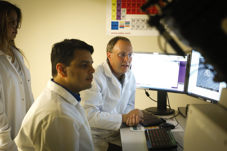
Housed in the Baylor Sciences Building, the CMI is home to some of the most powerful, advanced and expensive scientific instruments on campus, and is a key ingredient in the University’s goal to achieve Research 1/Tier 1 recognition.
“Every Tier 1 university has certain core facilities, and an advanced microscopy core facility is one of them,” Dr. Bernd Zechmann, CMI director and associate research professor said. “For cutting-edge research at Tier 1 universities, you need cutting-edge microscopes and related expertise, and the CMI offers that to all faculty so they can implement these technologies in their research.”
These and other more conventional scientific instruments are kept busy with approximately 80 active users coming from nine academic departments and 36 research groups. There are groups outside campus which collaborate with Baylor and make use of the CMI’s resources as well, including the U.S. Department of Defense, pharmaceutical companies, various businesses, and universities and research institutes in the United States and Europe.
Significant Investment
The CMI at Baylor opened in 2014 with Zechmann as its first director. Since then, more than $3.5 million has been invested in new equipment and a new lab that opened in May 2020. Having all the instruments in one location facilitates access, training and maintenance.
“High-end microscopes can cost anywhere between $150,000 and $10 million and cannot be fully used or maintained by a single professor or research group,” Zechmann said. “Thus, Baylor buys high-end microscopes and places them in the CMI, which are then available for multiple users.”
“For cutting edge research at Tier 1 universities, you need cutting-edge microscopes and related expertise, and the CMI offers that to all faculty so they can implement these technologies in their research.”Dr. Bernd Zachmann
Zechmann said some instruments have been donated to the CMI by retiring faculty, and some were obtained through funds that come with research grants. A new $6 million transmission electron microscope is expected to be installed in the spring of 2022. It is the first instrument of its type in the United States, and with the ability to image atoms at a resolution of 0.1 nm, it will be what Zechmann terms the “flagship” of the CMI.
Despite the high cost and complexity of these instruments, they can be used by anyone with proper training, available through group courses or personal instruction.
“I try to come up with simple standard operation procedures and train students and faculty so they can use these microscopes independently as much as possible,” Zechmann said. “If a student is dedicated and works full time on a specific project with a specific microscope, he or she can become fully independent within two to four weeks.”
Student Involvement
Dr. Marina George used the resources of the CMI as she earned her PhD from Baylor in environmental science with a focus on nanotoxicology — which studies how tiny, nano-size materials affect health. She currently is working at a U.S. Air Force research lab in Ohio where she is looking at how to detect molecular biosignatures of stress created by the extreme temperatures endured by airmen.
George’s master’s thesis involved the characterization of materials, which required her to learn how to use a variety of instruments in the CMI. The proficiency she developed with the equipment led her to be the teaching assistant in the lab for a year.
“Working in the CMI taught me about research in general and how you can take different techniques and apply them in different ways,” George said. “I hope I can eventually have my own microscopy lab or run a center or work for a national lab. My Baylor experience definitely influenced and positively impacted that.”
Grace Aquino is completing her PhD in environmental science at Baylor with an emphasis on human health and toxicology. Her research in the CMI explores how atmospheric pollution such as diesel exhaust particles affect the blood-brain barrier. She’s also trying to discover how microglial cells found in the central nervous system defend the brain from viruses, bacteria and chemicals, but at the same time block medicines that treat neuro-degenerative diseases.
Aquino would like to continue developing her testing models in postdoctorate study, or perhaps go directly into industry.
“One of the appeals of the model I’m developing is that you can use it for testing drug candidates to see which ones cross the blood-brain barrier and which ones might not,” Aquino said. “If they’re interested in that model or that sort of line of neurotoxicity and neuropharmaceuticals, I have a skill set that may be applicable.”
This is the first in a series of stories examining the purpose and contributions of the academic centers and institutes within the Baylor University College of Arts & Sciences.
